
Figure 2.
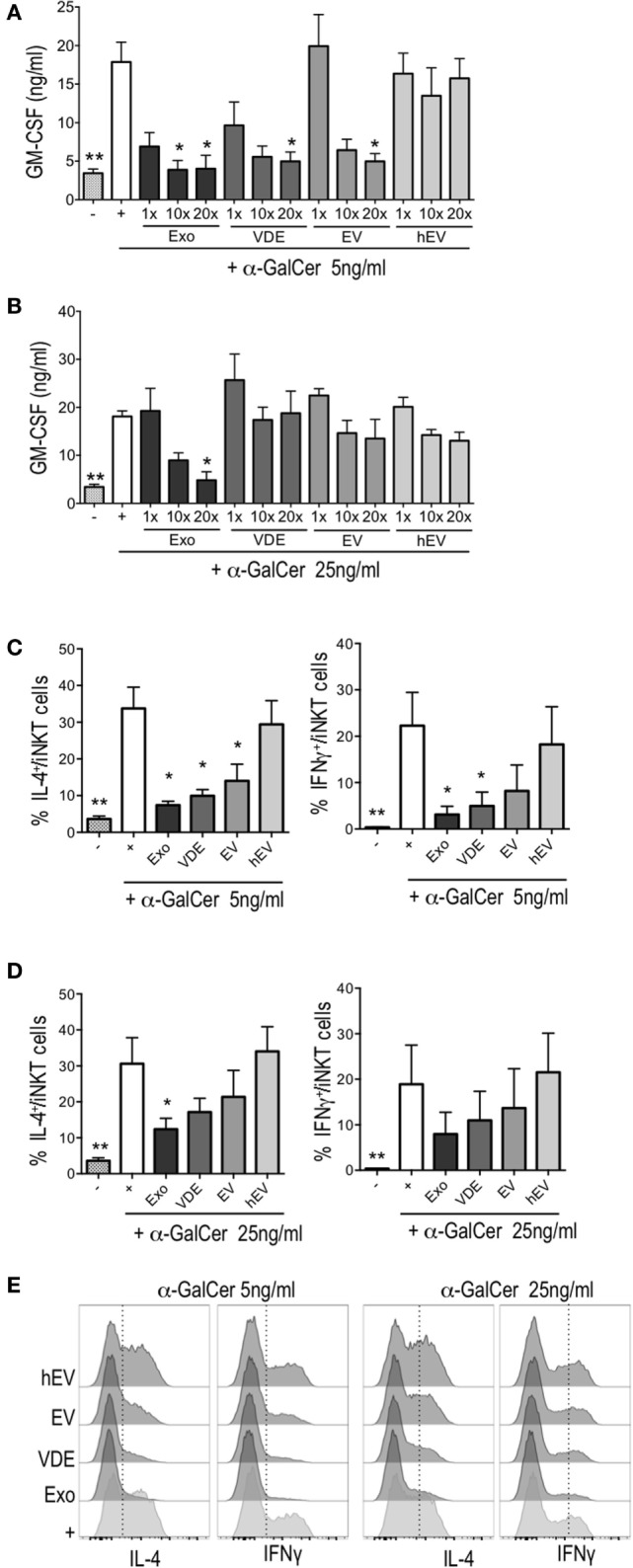
Leishmania infantum Exo, extracellular vesicle (EV), and vesicle-depleted-exoproduct (VDE) altered invariant natural killer T (iNKT) cell function. The polyclonal human iNKT cell line was incubated with CD1d-transfected C1R cells alone (−) or previously loaded with α-GalCer 5 ng/ml (A,C) or 25 ng/ml (B,D) with (+) or without L. infantum Exo, EV, VDE, or hEV. iNKT cell activation was measured by assessing GM-CSF concentrations in culture supernatants (A,B) and by determining the percentage of IL-4+ or IFNγ+ cells among the iNKT cell line (C–E). Data show means ± SEM and the results are representative of two to three independent experiments (n = 4). All groups were tested versus the positive α-GalCer (+) control group. *p < 0.05, **p < 0.01. (E) Representative FACS profile of the results presented at (C,D).