
Figure 4.
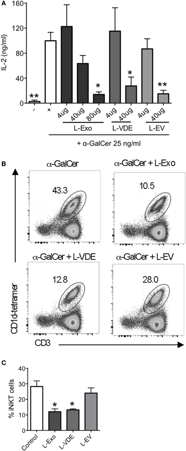
Lipids from Leishmania infantum Exo, extracellular vesicle (EV), and vesicle-depleted-exoproduct (VDE) mimicked the inhibition of invariant natural killer T (iNKT) cell activation. (A) Plate-bound mouse CD1d was loaded with α-GalCer (25 ng/ml) solely or in the presence of distinct doses of lipid extracts from L. infantum Exo, EV, VDE. 24.8 iNKT cell hybridoma was added and IL-2 production in the supernatants was measured. Data are expressed as means ± SEM of IL-2 concentrations in culture supernatants. Data are representative of two independent experiments (n = 4). All groups were tested versus the positive α-GalCer (+) control group. *p < 0.05, **p < 0.01. (B) Representative FACS profile showing the percentage of iNKT cells obtained after culture of peripheral blood mononuclear cell with α-GalCer in the presence of total lipid extracts from L. infantum Exo, EV, VDE. (C) Histograms represent the percentage of iNKT cells, as described in (B). Data are expressed as means ± SEM and they are representative of two to five independent experiments.