
Figure 3.
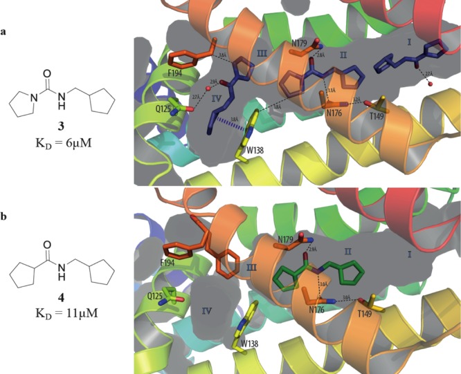
(a) X-ray crystal structure of urea 3 (PDB code 5IOY) bound to EthR. Three units of ligand 3 soak inside the binding cavity of the protein, filling subpockets I, II, and IV. (b) X-ray crystal structure of amide 4 (PDB code 5IOZ) bound to subpocket II of EthR.