
Figure 6.
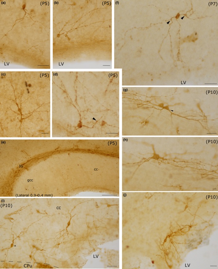
Photomicrographs of intracallosal NK1IP ‐n at three postnatal ages (P5‐P7‐P10). a–e: P5; f: P7; g–j: P10. a–d: Four intracallosal NK1IP ‐n showing different morphologies. (a) An ovoid NK1IP ‐n with a thick principal dendrite directed toward the ependymal cc region. (b and d) Two round NK1IP ‐n close to the ependymal cc region, whose dendrites are directed toward the dorsal, posterior and anterior cc regions. (c) Two adjacent NK1IP ‐n in the middle of the cc. (e) Medial cc region, probably between lateral at 0.9 and 0.4 mm (comparable with those of the adult): no neurons are found at this cc level. Several NK1IP ‐n are visible over the cc, in the IG. (f) An ovoid NK1IP ‐n with dendrites directed in all directions, including the ependymal cc region. Arrowhead: a growth bud. (g) Polygonal NK1IP ‐n. (i) Several intracallosal NK1IP ‐n. A neuron (asterisk) sends its dendrites into the CPu. (j) A bipolar NK1IP ‐n close to the ependymal region of the cc. Arrowheads in d and f indicate growth buds at branching points. Calibration bars: 25 μm in a–d, f, g, i, j; 100 μm in e; 10 μm in h