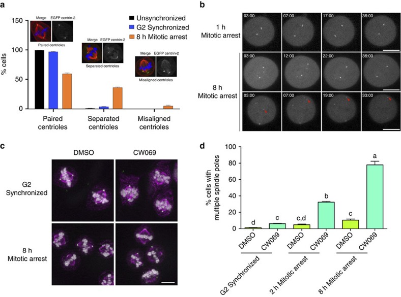
Figure 2. Spindle pole integrity and spindle bipolarity following mitotic delay is maintained by HSET-mediated centriole clustering.
(a) Representative phenotypes observed for centriole pairs in unsychronized, G2-synchronized and prometaphase-arrested cells. Error bars represent s.e.m. for three replicate experiments, with 300 cells scored per condition per experiment. (b) 4D time-lapse microscopy of eGFP centrin-2-expressing cells following monastrol washout to allow bipolar spindle assembly. Image stacks were acquired every minute, beginning ∼3 min following monastrol washout. Red arrow denotes the long-distance clustering of an individual centriole. Scale bar, 10 μm. Also see Supplementary Movies 1–3. (c) G2-synchronized or 8 h mitotically arrested RPE1 cells were allowed to progress into metaphase for 30 min in the presence of 0.1% DMSO or 350 μM CW069 (HSET inhibitor), and then fixed and probed for α-tubulin (magenta), PCNT (green) and DNA (white). Scale bar, 10 μm. (d) Quantification of the frequency of multipolar spindles. Error bars represent s.e.m. from three experimental replicates, with 300 cells scored per condition per experiment. Data were arcsin-square root transformed to achieve a normal distribution. A two-factor ANOVA was performed with a Tukey–Kramer post hoc test to discern differences among individual means, with significant differences indicated with different lower-case letters.

