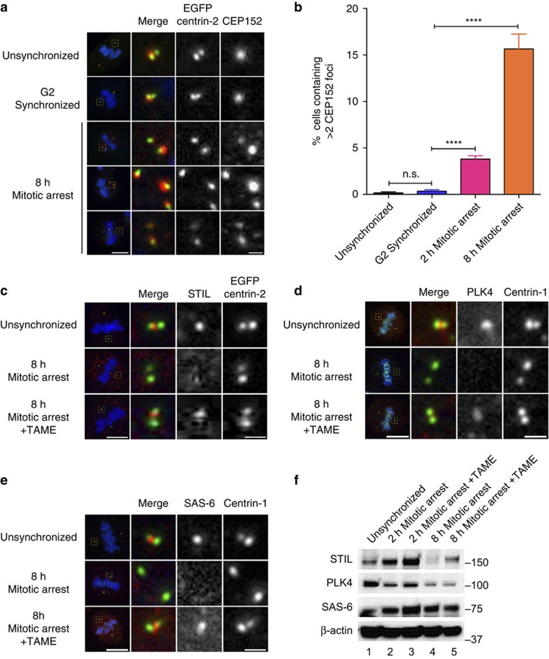
Figure 4. Loss of procentriolar markers during mitotic delay.
(a) CEP152/Asl localization in unsynchronized, G2-synchronized and cells subjected to mitotic delay. Lower left bar, 10 μm; Lower right bar, 1 μm. (b) Quantification of CEP152/Asl foci. Error bars represent s.e.m. for three replicate experiments, 300 cells scored per condition per experiment, with significance determined by one-way ANOVA with Tukey–Kramer post hoc test, ****P≤0.0001. (c–e) Mitotic cells from unsynchronized cultures, or 8 h of mitotic arrest in the absence or presence of TAME. Cells were then fixed and probed for the presence of STIL (c), PLK4 (d) or SAS-6 (e). Lower left bars, 10 μm; Lower right bars, 1 μm. (f) Total protein levels of procentriole markers in cells treated in the conditions shown in c–e. See Supplementary Fig. 4 for quantification.

