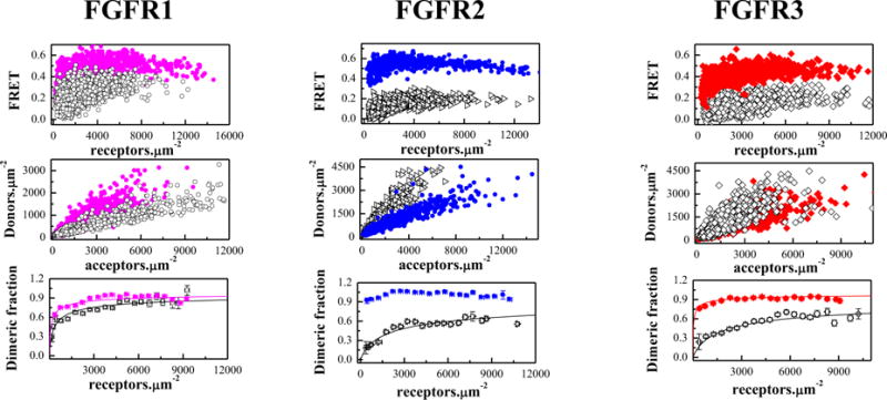
Figure 2.

The cysteine mutations stabilize truncated EC+TM FGFR dimers in the absence of ligands. Solid magenta circles: C178R FGFR1; Solid blue diamonds: C342R FGFR2; Solid red squares: C228R FGFR3. Top panels: FRET efficiencies for the mutants (solid color symbols) and the wild-types (open black symbols,8). Middle panels: Donor concentration versus acceptor concentration in each vesicle. Bottom panels: Dimeric fraction as a function of total receptor concentration (donors+acceptors). The QI-FRET method, used for data collection and analysis, has been described in detail in refs. 34,35,50
