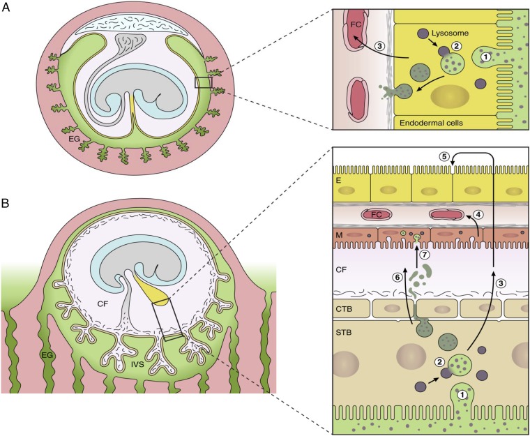
Fig. 7.
Diagrammatic comparison of the nutrient pathway during early pregnancy in the mouse (A) and the speculated pathway in the human (B). In the mouse, histotrophic secretions (green) released from the endometrial glands are phagocytosed (step 1) by the endodermal cells of the visceral layer of the inverted yolk sac. Following fusion with lysosomes (step 2), digestion of maternal proteins leads to release of amino acids that are transported (step 3) to the fetal circulation (FC). In the human, histotrophic secretions are released from the endometrial glands through the developing basal plate of the placenta into the intervillous space (IVS) and are phagocytosed (step 1) by the syncytiotrophoblast (STB) (42). We speculate that following digestion by lysosomal enzymes (step 2), free amino acids are transported (step 3) by efflux transporters to the coelomic fluid (CF), where they accumulate. Nutrients in the CF may be taken up by the mesothelial cells (M) of the yolk sac and transported (step 4) into the fetal circulation (FC). Alternatively, they may diffuse into the cavity of the yolk sac and be taken up by the endodermal cells (step 5). Some intact maternal proteins may also be released into the CF by exocytosis of residual bodies (step 6) and may be engulfed by the mesothelial cells (step 7). CTB, cytotrophoblast cells.

