Fig. 3.
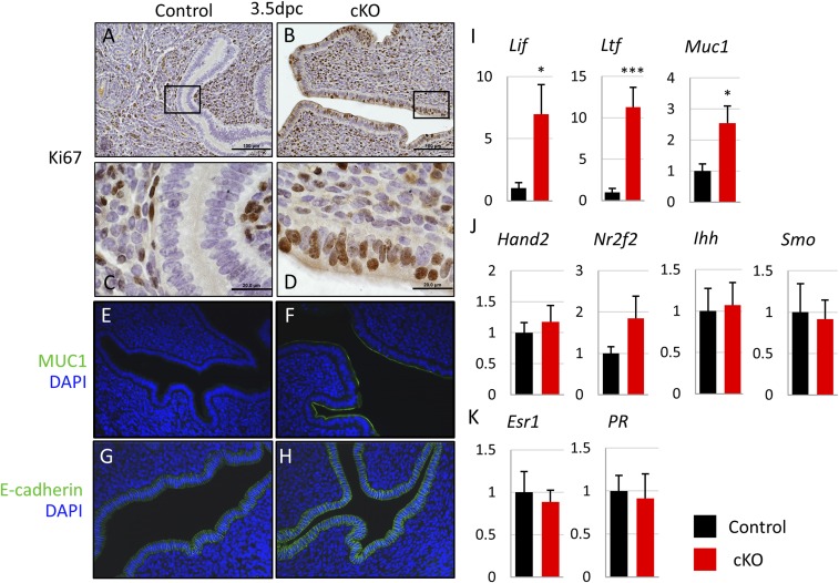
Fst-cKO mice fail to achieve uterine receptivity at 3.5 dpc. (A–D) Immunohistochemistry for the proliferation marker Ki67 with paraffin sections from pregnant control and Fst-cKO females at 3.5 dpc. Sections are counterstained with hematoxylin. Brown staining denotes Ki67+ proliferating cells. (C and D) Higher magnification of the boxed sections in A and B. (Scale bars: A and B, 100 μm; C and D, 20 μm.) (E and F) Immunofluorescence for the uterine receptivity marker MUC1 (green) and nuclear marker DAPI (blue). Expression of MUC1 in the epithelium at 3.5 dpc indicates a nonreceptive uterus. (G and H) Immunofluorescence for the uterine receptivity marker E-cadherin (green) and nuclear marker DAPI (blue). Expression of E-cadherin in the epithelium at 3.5 dpc indicates intact apical–basal polarity and a nonreceptive uterus. (Magnification: 20×.) (I–K) Relative expression of mRNA for E2- (I) and P4- (J) regulated genes and hormone receptors (K) in control (n = 3) and Fst-cKO (n = 6) females at 3.5 dpc. Uterine luminal epithelium and stroma were separated and purified after trypsin digestion. Lif, Ltf, Muc1, and Ihh are epithelial-expressed genes; Hand2, Nr2f2, Smo, Esr1, and PR are stromal-expressed genes. Gene-expression data are normalized to levels of 36B4 mRNA with the average value of the control set to one and are compared across genotype. Data are presented as mean ± SEM; *P < 0.05, ***P < 0.005.

