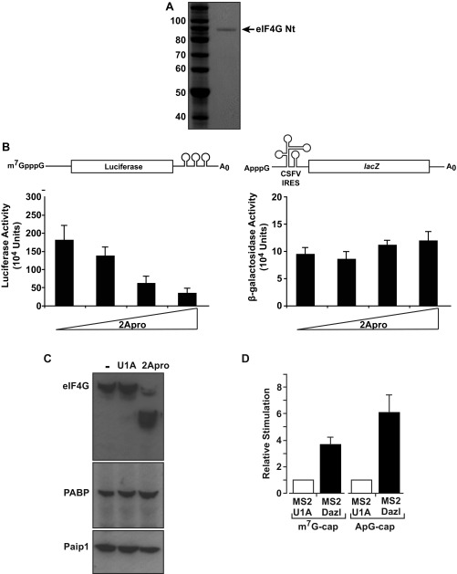
Fig. S6.
(A) Purified eIF4G. FLAG fusion of eIF4G (amino acids 1–532) visualized with Gelcode Blue. (B) 2A protease does not disrupt CSFV IRES-dependent translation initiation. Oocytes were injected with m7G-Luc-MS23 (Left) or CSFV–β-gal (Right) mRNAs (Fig. S2A, [3] and [8]) and increasing amounts of mRNA expressing 2Apro. Luciferase and β-gal activities were determined, showing that 2Apro inhibits cap-dependent (Left) but not eIF4G-independent (Right) CSFV IRES-driven translation, as expected. (C) Oocytes were uninjected (−) or injected with mRNA expressing 2Apro or U1A (a proteolytically and translationally inert control protein). After 3 h, extracts were prepared and endogenous eIF4G, PABP, and Paip1 were analyzed by immunoblotting. (D) Dazl activates a nonphysiologically (ApppG)-capped reporter mRNA in a tether-function assay. Oocytes expressing MS2-U1A or MS2-Dazl were coinjected with β-gal mRNA and m7G-Luc-MS23 or ApG-Luc-MS23 (Fig. S2A, [1], [2], and [7]). Effects on translation were measured by luciferase assay normalized for β-gal activity. Translational stimulation relative to MS2-U1A (set to 1) is plotted. Error bars indicate SEM from six repeats.

