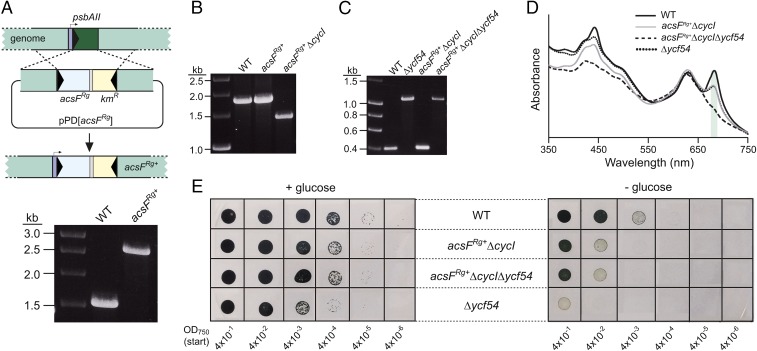
Fig. 2.
Construction and phenotypic analyses of Synechocystis cyclase mutants. (A) Diagram depicting replacement of the psbAII gene with acsFRg via pPD[acsFRg] (Upper), and construction of the fully segregated strain confirmed by colony PCR (Lower). (B and C) Inactivation of cycI (B) and ycf54 (C) genes via replacement with chloramphenicol and zeocin resistance cassettes, respectively, confirmed by colony PCR. (D) Whole-cell absorption spectra of strains grown mixotrophically under low light conditions. The peaks for Chl-containing complexes are marked with a green shadow. (E) Drop growth assays of strains on solid agar, supplemented with or lacking glucose. Photographs were taken after incubation for 12 d.

