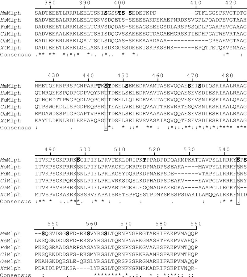
Fig. S1.
Alignment of Mlph ABDs from mouse (Mm), human (Hs), Fukomys (Fd), dog (Cl), sheep (Oa), and Xenopus (Xt). Sequence alignment of selected Mlph ABDs revealed numerous conserved serine/threonine residues that represent potential PKA targets. Serine and threonine residues that were found to be phosphorylated in vivo are in bold. Boxes indicate conserved phosphorylatable serine residues. Numbers represent the residue numbers according to MmMlph. Asterisks, colons, and dots indicate positions that are fully, partially, or weakly conserved, respectively.

