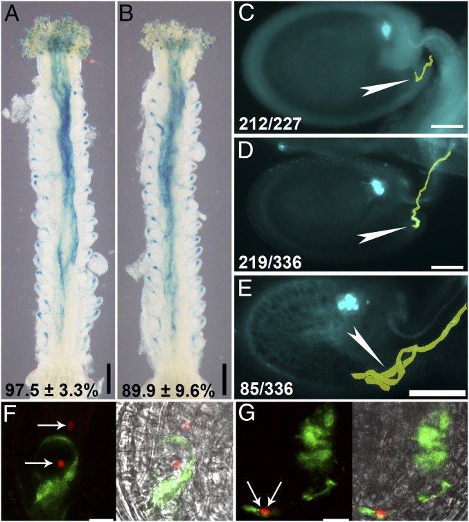
Fig. 2.
Female gametophytes of ap1g1 g2 are compromised in pollen tube reception. (A and B) Representative histochemical GUS analysis of wild-type (A) or ap1g1+/− g2 (B) pistils emasculated and hand-pollinated with ProLAT52:GUS pollen at 12 HAP. Results shown at the bottom are means ± SD, n = 25. (C–E) Representative aniline blue-stained WT ovule (C), ap1g1+/− g2 ovule with normal pollen tube reception (D), ap1g1+/− g2 ovule with a tangled pollen tube (E) at 48 HAP. Pollen tubes are pseudocolored in yellow. Arrowheads point at the micropyle. At the bottom: displayed ovules/all ovules examined. (F and G) CLSM of a wild-type (F) or ap1g1 g2 (G) embryo sac invaded by a ProLAT52:GFP;ProHTR10:HTR10-mRFP pollen tube. The right images are merges of GFP, RFP, and BF channels. Arrows in F and G indicate the position of sperm cells. (Scale bars: A and B, 200 µm; C–E, 50 µm; F and G, 10 µm.)

