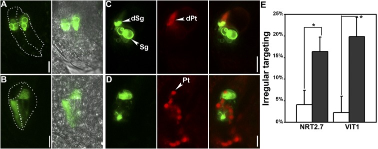
Fig. S4.
Targeting of tonoplast proteins is interfered in synergids by AP1G loss of function. (A–D) CLSM of ProDD39:VIT1-GFP in synergids of WT (A and C) or ap1g1/+ g2 (B and D). Pistils from ProDD39:VIT1-GFP or ProDD39:VIT1-GFP;ap1g1/+ g2 plants were either emasculated at maturation for visualization (A and B) or emasculated and pollinated with ProLAT52:DsRed pollen and visualized at 8 HAP (C and D). Merges of GFP and RFP channels are shown at Right. Dotted lines illustrate synergid cells. dPt, discharged pollen tube; dSg, degenerating synergid; Pt, pollen tube; Sg, synergid. (E) Percentage of ovules in which GFP-NRT2.7 and VIT1-GFP were not associated with the tonoplast in synergids. Results are means ± SEM of three independent experiments involving 150 ovules. Open columns indicate WT, whereas filled columns indicate ap1g1/+ g2. Asterisk indicates significant difference (t test, P < 0.01). (Scale bars: 10 µm.)

