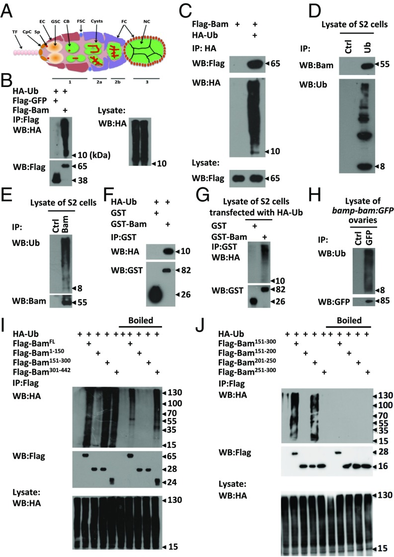
Fig. 1.
Bam is an ubiquitin-association protein. (A) Schematic diagram displaying the ovarian germarium with various cell types and organelles including the terminal filament (TF), cap cells (CpC), spectrosome (Sp), escort cells (EC), germ-line stem cells (GSC), cystoblasts (CB), follicle stem cells (FSC), cysts, follicle cells (FC), and Nurse cells (NC). (B and C) S2 cells were transfected with indicated plasmids. Cell lysates were subjected to immunoprecipitation assays using Flag beads (B) or anti-HA antibody (C), followed by Western blot assays. (D and E) Lysates of S2 cells were immunoprecipitated with anti-ubiquitin (D) or anti-Bam (E) or mouse IgG (control) antibody, followed by Western blot assays. (F and G) Purified HA-ubiquitin proteins (F) or lysates of S2 cells transfected with HA-Ub plasmids (G) were mixed with GST or GST-Bam, followed by immunoprecipitation and Western blot assays. (H) Ovaries of P{bamp-bam:GFP} were dissected and lysed, followed by immunoprecipitation using anti-GFP or mouse IgG (control) antibody. Western blot assays were performed to detect the levels of indicated proteins. (I and J) S2 cells were transfected with indicated plasmids. Cell lysates were subjected to immunoprecipitation by using Flag beads with or without boiling pretreatment as indicated. Western blot assays were performed to detect the HA signal. All of the biochemical experiments were performed at least three times.

