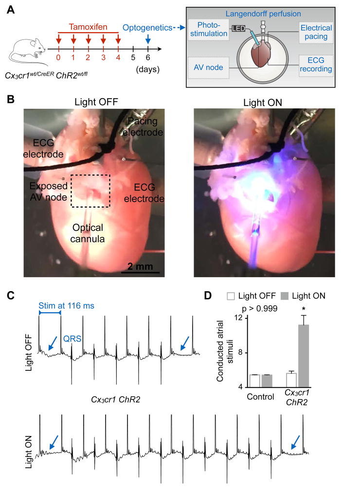
Figure 5. Optogenetics Stimulation of AV Node Macrophages Improves Nodal Conduction.
(A) Experimental outline. Hearts of Cx3cr1wt/CreER (control) or tamoxifen-treated Cx3cr1wt/CreER ChR2wt/fl (Cx3cr1 ChR2) mice were perfused in a Langendorff setup. Recording and pacing electrodes were connected to the heart and illumination with a fiber optic cannula was focused on the AV node.
(B) Images illustrating the optogenetics experimental setup during a light off and on cycle.
(C) Representative ECG recordings from a Cx3cr1 ChR2 heart illustrating the number of conducted atrial stimuli between two non-conducted impulses of a Wenckebach period during light off and on cycles. Arrows indicate failure of conduction leading to missing QRS complexes. Stim, stimulation.
(D) Representative bar graphs of a control and Cx3cr1 ChR2 heart showing the number of conducted atrial stimuli between two non-conducted impulses of a Wenckebach period during light off and on cycles. Data are mean ± SEM, *p < 0.05, Kruskal-Wallis test followed by Dunn’s posttest.
See also Figure S5.

