
Figure 4. Vitamin A deficiency disrupts tissue resident macrophages.
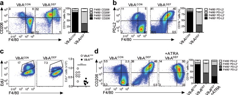
Peritoneal macrophages were evaluated from either vitamin A deficient (Vit-ADEF) mice or control (Vit-ACON) mice after treatment with IL-4c or without treatment. Representative FACS plots for (a) Flow cytometry analysis of peritoneal macrophage Vit-ADEF(n=7) and Vit-ACON(n=7) mice. Stacked bar graph shows increased frequencies of F4/80+ CD206+and F4/80+ CD206−expressing CD11b+populations; and (b) Flow cytometry analysis of peritoneal macrophages from Vit-ADEF(n=5) and Vit-ACON(n=5) mice. Stacked bar graph shows increased frequencies PD-L2 and F4/80 expression. (c) EdU incorporation from peritoneal macrophages of IL-4c treatedVit-ADEF(n=8) and Vit-ACON(n=6) mice. (d) Vit-ADEFmice were treated with all-trans RA every 2 days for 14 days prior to IL-4c treatment. Representative FACS plots of PD-L2 expression in peritoneal macrophages of Vit-ACON(n=5), Vit-ADEF(n=5), and Vit-ADEFmice treated with all-trans RA (ATRA) (n=5). Stacked bar graph indicates frequencies of F4/80 and PD-L2 in CD11b+ cells. **P < 0.01. Unpaired Students T-test.