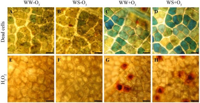
FIGURE 1.

Localization of dead cells visualized with Evans blue staining (A–D) and of hydrogen peroxide (H2O2) visualized the 3,3′-diaminobenzidine (DAB) uptake method (E-H) in Quercus ilex leaves (i) well-watered (WW) and exposed to charcoal filtered air (WW-O3); (ii) water stressed (20% of effective evapotranspiration daily for 15 days) and exposed to charcoal filtered air (WS-O3); (iii) well-watered and exposed to acute ozone (200 nL L-1 for 5 h) (WW+O3); (iv) water stressed and O3 fumigated (WS+O3). The assays were performed 96 h FBE. Bars 50 μm.
