Abstract
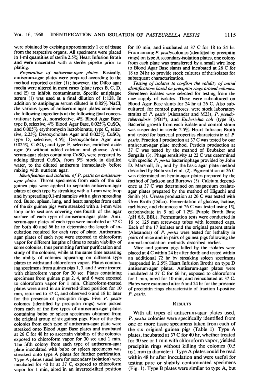
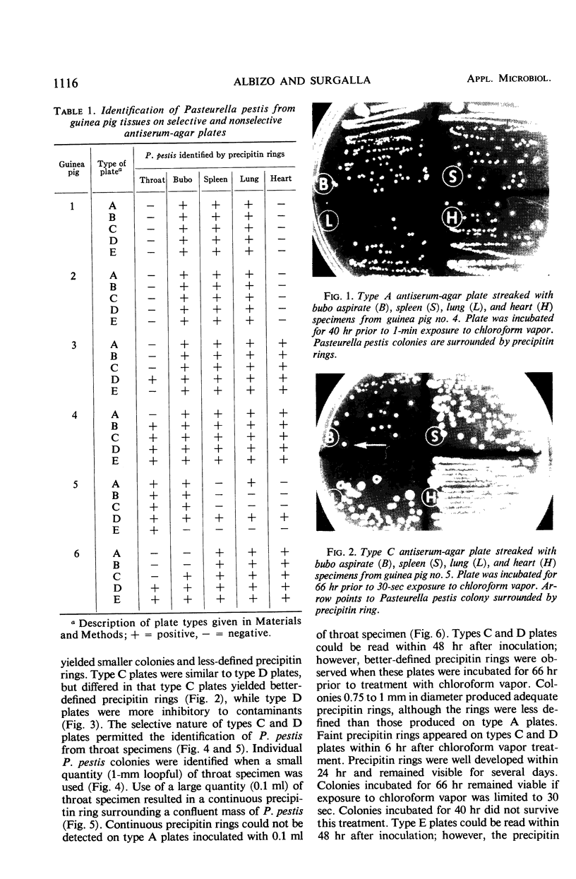
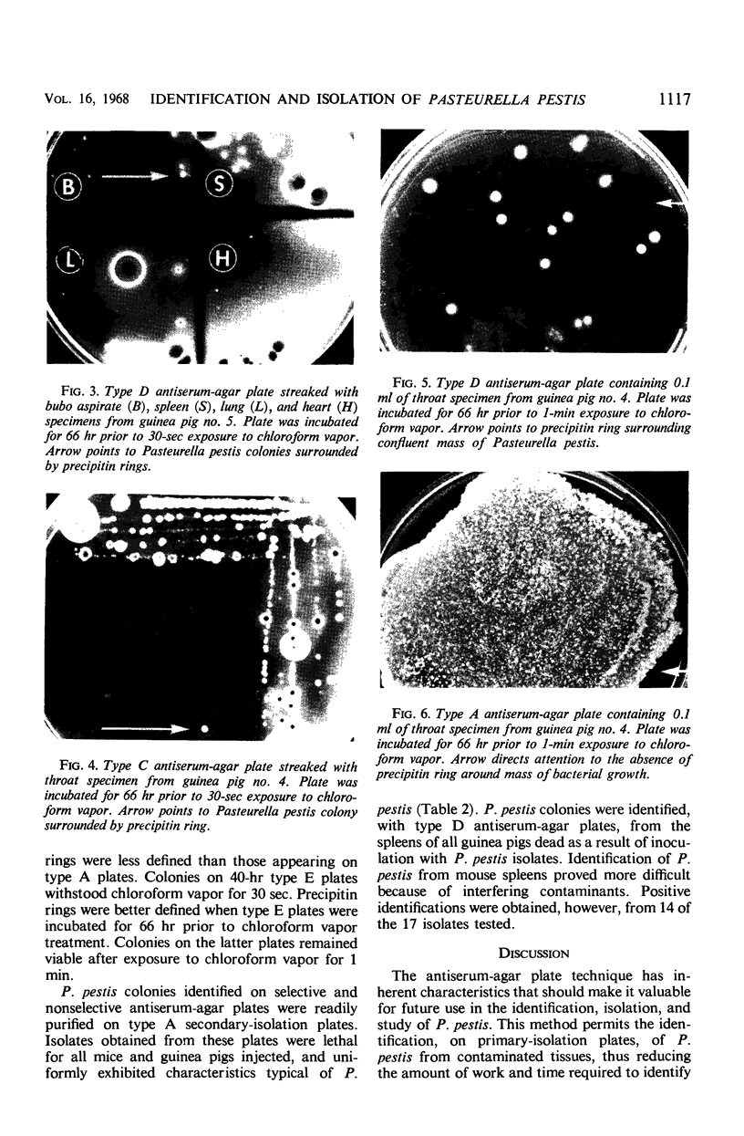
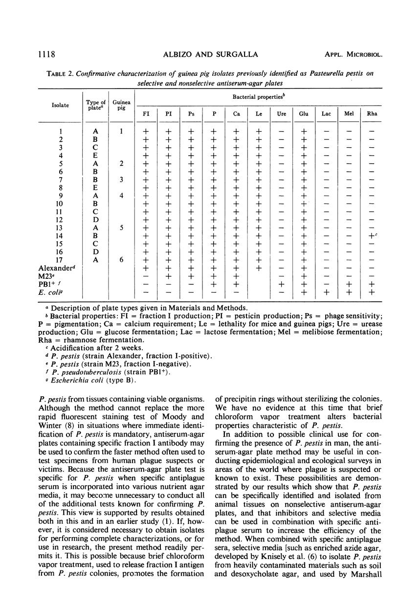
Pasteurella pestis colonies were specifically identified on antiserum-agar plates used for primary culture of tissues from experimentally infected guinea pigs. Both selective and nonselective antiserum-agar plates were used to identify P. pestis from guinea pigs kept at 22 C for periods up to 4 days after death from plague. Colonies identified as P. pestis on selective and nonselective antiserum-agar plates, by the appearance of precipitin rings following brief chloroform vapor treatment, remained viable and were subsequently purified on nonselective antiserum-agar plates. Isolates obtained in this manner were uniformly lethal when injected into mice and guinea pigs, and conformed to standard laboratory criteria for P. pestis. P. pestis was identified on selective antiserum-agar plates from the spleens of all guinea pigs killed by the isolates, and from a large majority of the mice. The practical value and confirmative nature of the method were demonstrated.
Full text
PDF
Images in this article
Selected References
These references are in PubMed. This may not be the complete list of references from this article.
- Albizo J. M., Surgalla M. J. Specific identification of fraction I-positive Pasteurella pestis colonies on antiserum-agar plates. Appl Microbiol. 1968 Apr;16(4):649–655. doi: 10.21236/ad0835494. [DOI] [PMC free article] [PubMed] [Google Scholar]
- BALTAZARD M., DAVIS D. H., DEVIGNAT R., GIRARD G., GOHAR M. A., KARTMAN L., MEYER K. F., PARKER M. T., POLLITZER R., PRINCE F. M. Recommended laboratory methods for the diagnosis of plague. Bull World Health Organ. 1956;14(3):457–509. [PMC free article] [PubMed] [Google Scholar]
- BRUBAKER R. R., SURGALLA M. J. Pesticins. II. Production of pesticin I and II. J Bacteriol. 1962 Sep;84:539–545. doi: 10.1128/jb.84.3.539-545.1962. [DOI] [PMC free article] [PubMed] [Google Scholar]
- BURROWS T. W., JACKSON S. The pigmentation of Pasteurella pestis on a defined medium containing haemin. Br J Exp Pathol. 1956 Dec;37(6):570–576. [PMC free article] [PubMed] [Google Scholar]
- HIGUCHI K., SMITH J. L. Studies on the nutrition and physiology of Pasteurella pestis. VI. A differential plating medium for the estimation of the mutation rate to avirulence. J Bacteriol. 1961 Apr;81:605–608. doi: 10.1128/jb.81.4.605-608.1961. [DOI] [PMC free article] [PubMed] [Google Scholar]
- KNISELY R. F., SWANEY L. M., FRIEDLANDER H. SELECTIVE MEDIA FOR THE ISOLATION OF PASTEURELLA PESTIS. J Bacteriol. 1964 Aug;88:491–496. doi: 10.1128/jb.88.2.491-496.1964. [DOI] [PMC free article] [PubMed] [Google Scholar]
- MOODY M. D., WINTER C. C. Rapid identification of Pasteurella pestis with fluorescent antibody. III. Staining Pasteurella pestis in tissue impression smears. J Infect Dis. 1959 May-Jun;104(3):288–294. doi: 10.1093/infdis/104.3.288. [DOI] [PubMed] [Google Scholar]
- Marshall J. D., Jr, Quy D. V., Gibson F. L. Asymptomatic pharyngeal plague infection in Vietnam. Am J Trop Med Hyg. 1967 Mar;16(2):175–177. doi: 10.4269/ajtmh.1967.16.175. [DOI] [PubMed] [Google Scholar]


