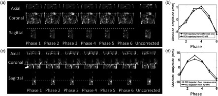
Fig. 7.
(a) Six-phase 4-D-MRI images, with uncorrected images as comparison and (b) comparison of ROI motion trajectories between 4-D-MRI and reference cine MR for a representative healthy volunteer. (c) Six-phase 4-D-MRI images (white arrow points out a cyst in liver), with uncorrected images as comparison and (d) comparison of ROI motion trajectories between 4-D-MRI and reference cine MR for a representative cancer patient. A benign cyst in the liver is pointed out by the white arrow.

