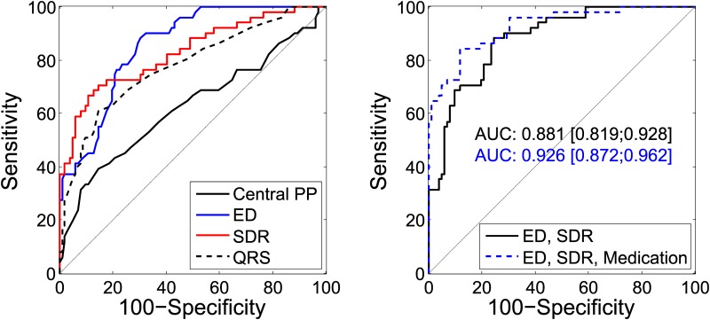
Fig 2. ROC curve analysis.
Left: Comparison of the ROC curves obtained with central pulse pressure (PP), ejection duration (ED), S to D ratio (SDR) and QRS-duration. Right: ROC curves obtained with a combination of ED and SDR, and ED and SDR when adjusted for medication. Area under the curve (AUC) and 95% confidence interval are given in the same colors.

