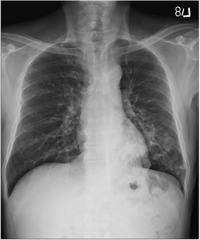
Fig 1. Primary pulmonary tuberculosis pattern with lower lung field lesions.
A 53-year-old male with culture-proven pulmonary tuberculosis and a history of diabetes mellitus. The patient had a glycosylated hemoglobin (HbA1c) of 13.5%. CXR revealed patchy infiltrates and ill-defined acinar shadows in the left lower lung field.

