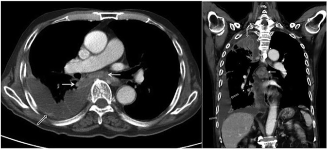
Fig 3. Primary pulmonary tuberculosis pattern with lymphadenopathy and pleural effusion.
A 94-year-old male with culture-proven pulmonary tuberculosis and a history of diabetes mellitus. The patient had a glycosylated hemoglobin (HbA1c) of 9.4%. Axial (A) and coronal (B) contrast-enhanced thoracic CT scans revealed loculated right pleural effusion with pleural thickening (open arrows) and enlarged subcarinal and right interlobar lymph nodes with central low attenuation and peripheral rim enhancement (arrows).

