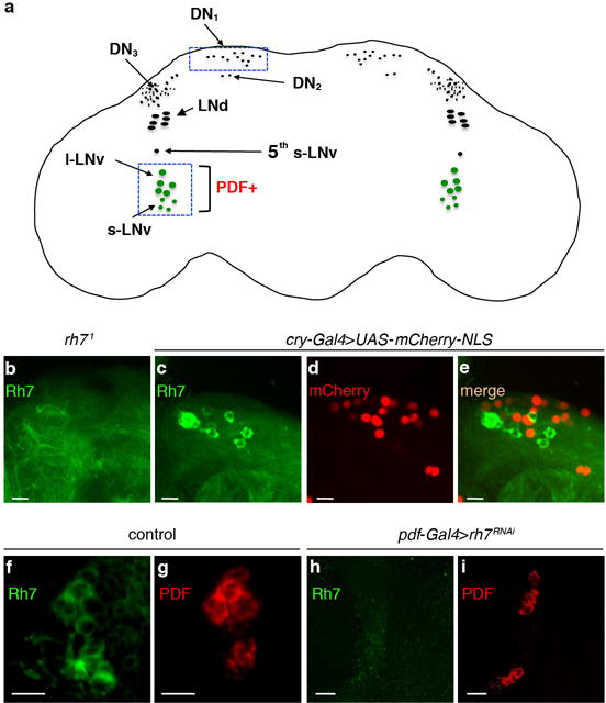
Extended Data Figure 3. Expression of Rh7 in non-clock neurons located in the dorsal part of the brain.

a, Cartoon of a fly brain showing different groups of clock neurons. The boxed areas indicate locations of two groups of Rh7-positive cells. b, rh71 brain stained with anti-Rh7. c–e, Double labeling of the dorsal region of the brain with a clock neuron reporter (cry-Gal4E13>UAS-mCherry-NLS) and anti-Rh7. The scale bars in b–e indicate 20 μm. c, Anti-Rh7. d, Anti-mCherry. e, Merge of c and d. f, Control brain stained with anti-Rh7. g, Control brain stained with anti-PDF. h, pdf-Gal4>rh7RNAi brain stained with anti-Rh7. i, pdf-Gal4>rh7RNAi brain stained with anti-PDF. The scale bars in f–i indicate 10 μm.
