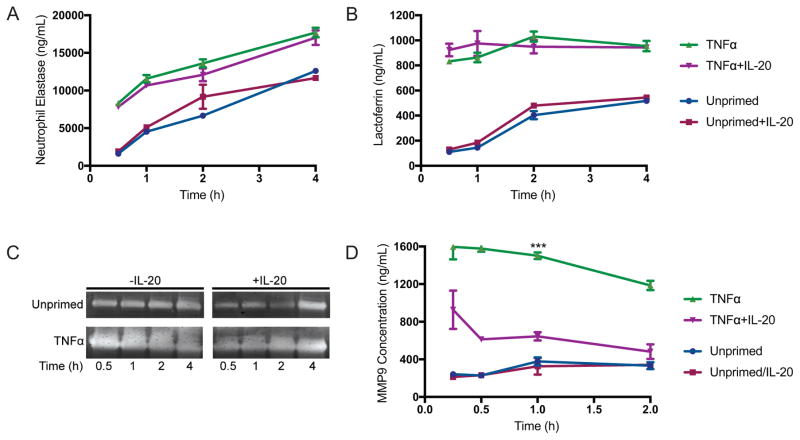
Figure 3. IL-20 inhibits exocytosis of tertiary (gelatinase) granules.
(A) Concentration of neutrophil elastase (primary/azurophilic granule marker) in supernatants from human neutrophils, incubated with S. aureus MOI 1 for indicated times, measured by reaction with colorimetric substrate, calculated using standard of known concentration. (B) Lactoferrin concentration, marker for secondary (specific) granules, measured by ELISA of neutrophil supernatants. Data shown in (A) and (B) as mean +/− SEM from three healthy donors. (C) Gelatin zymography of supernatants from neutrophils infected for indicated times with S. aureus (MOI 1). Results shown are representative of neutrophils from four independent healthy donors. (D) Concentration of MMP9, marker for tertiary (gelatinase) granule release, in supernatants of neutrophils infected for indicated times with S. aureus (MOI 1) +/− IL-20. Data shown as mean +/− SEM from four healthy donors. ***p<0.001 compared to TNFα+IL-20-treated neutrophils by Two-way ANOVA.

