Figure 2.

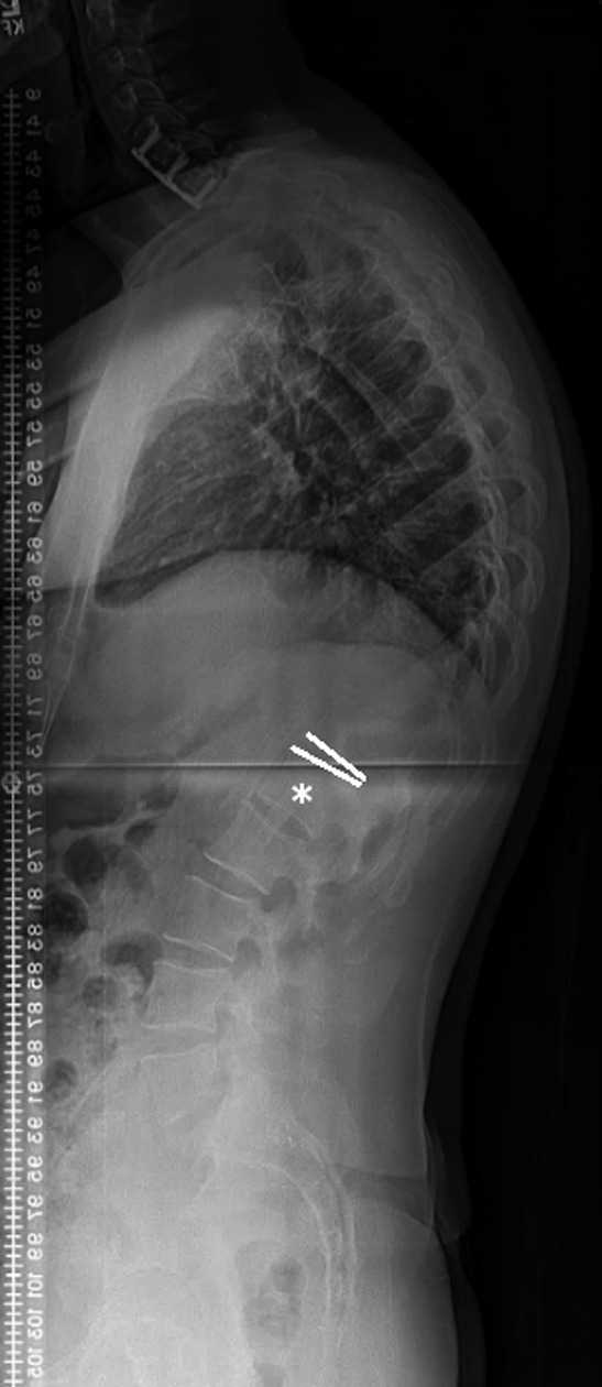
Lateral standing radiograph of a 33-year-old male with Scheuermann’s kyphosis. According to the first lordotic disc method, the distal fusion level (L1) is the vertebra subjacent to the FLD and marked by the “*” in this image.

Lateral standing radiograph of a 33-year-old male with Scheuermann’s kyphosis. According to the first lordotic disc method, the distal fusion level (L1) is the vertebra subjacent to the FLD and marked by the “*” in this image.