
Fig. 4.
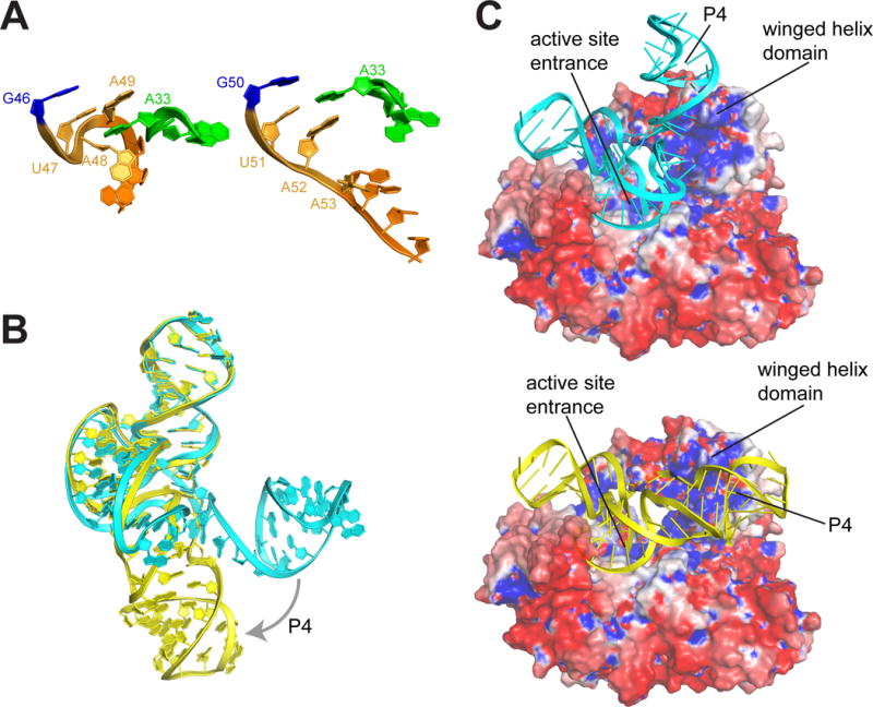
Model of ZIKV xrRNA-Xrn1 interaction. A. Comparison of the S4 region (orange) and adjacent in the partially folded MVE (left) and fully folded ZIKV (right) xrRNAs. B. Overlay of the MVE (cyan) and ZIKV (yellow) structures, showing the change in the position of the P4/L4 hairpin. C. Models of the MVE (top) and ZIKV (bottom) xrRNAs docked onto the surface of Xrn1 colored by electrostatic potential (blue = positive, red = negative). Structural features are labeled.