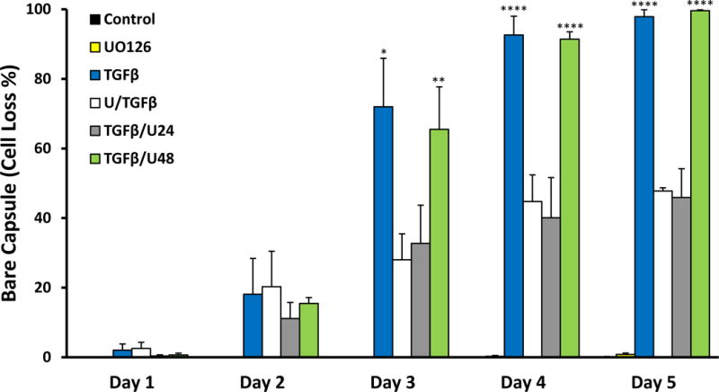
Figure 10.

Summary of cell loss in explants treated without TGFβ (control), with UO126, TGFβ, U/TGFβ, TGFβ/U24 and TGFβ/U48 over 5 days. Trends revealed that TGFβ and TGFβ/U48 had similar rates in cell loss. U/TGFβ and TGFβ/U24 exhibited a similar progression of cell loss (error bars represent SEM; ANOVA with Tukey’s post-hoc test was used to analyze data; *p<0.05, **p<0.01, ****p<0.0001; when compared to control group). Cell loss was expressed as a percentage of bare capsule appearance (i.e. 100% bare capsule corresponds to 100% cell loss).
