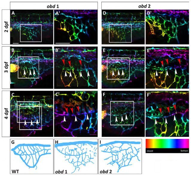
Fig. 6.
obd mutants have a variable SIVP pattern and SIVP overgrowth. Depth-coded confocal stacks of SIVP development in two individual embryos (A–C′ and D-F′) of obdfov01b; Tg(fli:EGFP)y1 line from 2 to 4 dpf showing variability in SIVP pattern. The border of the inner vascular basket is indicated by white arrowheads. Shared vessels between the inner and outer basket are indicated by red arrowheads. Scale bars represent 100 μm. (G–I) Schematics of SIVP phenotype in wild-type (Fig. 2C), obd 1 (Fig. 6C) and obd 2 (Fig. 6F) embryos at 4 dpf.

