
Figure 3. Visualization of microtubule sliding in Drosophila neurons using photoconvertible tubulin.
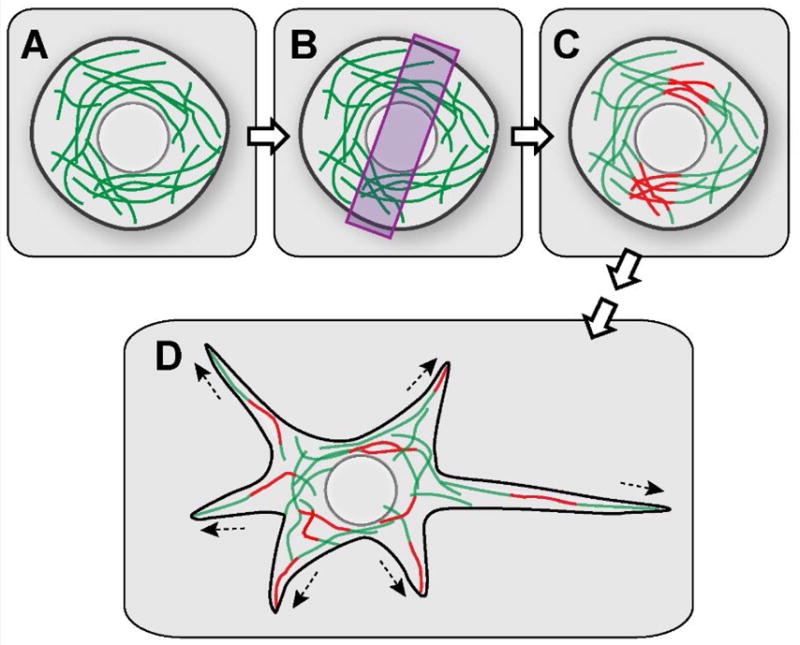
(A) A spherical-shaped young neuron expressing photoconvertible EOS-tagged α-tubulin. (B) 400nm light is applied to a restricted area to photoconvert a subset of microtubules in the young neurons. (C) A subset of microtubules is photoconverted from green to red. (D) Red microtubule fragments are scattered throughout the cell body and in the newly formed neurites by microtubule-microtubule sliding, revealed by time-lapse movies.