
Figure 2. Overview of picornaviral RdRP structures.
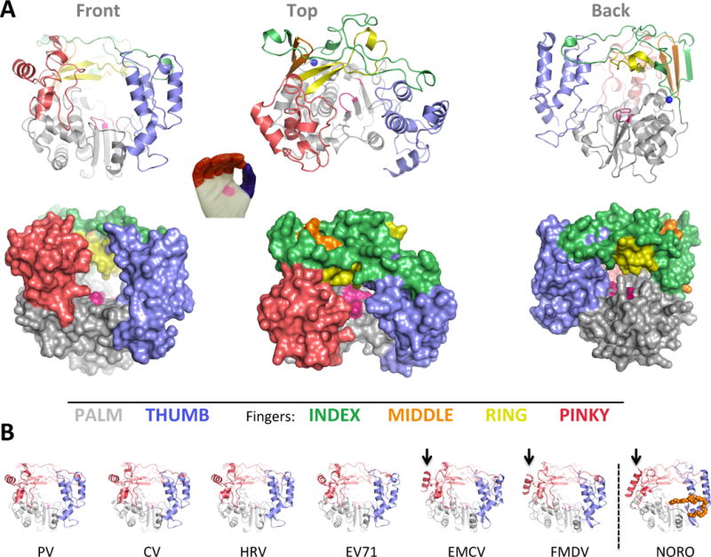
(A) Cartoon and surface representations of poliovirus 3Dpol in three different orientations. The structure resembles a cupped right hand composed of palm, fingers, and thumb domains. The fingers domain can be further divided into five distinct structures (per color key), and the active site in the palm domain is shown as a patch of magenta. Note that the index finger reaches across the palm to contact the top of the thumb, creating a channel at the back of the enzyme whereby NTPs access the active site. (B) All the picornaviral 3Dpol structures solved to date exhibit a very high degree of structural homology. Note that one helix on the pinky finger changes orientation between the enteroviruses and EMCV/FMDV groups, with the latter resembling the helix orientation seen in the non-picornaviral norovirus 3Dpol. Norovirus polymerase also has a C-terminal extension (orange) that reaches into the RNA exit channel.