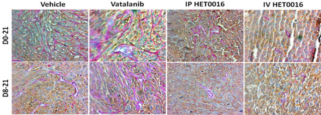
Figure 6. Vatalanib increased number of VM and novel IV formulation of HET0016 decreased VM of human GBM in rat model.
(A) Immunohistochemistry data showing PAS stain representing increased VM (pink vessel-like strictures due to PAS+) in Vatalanib treated tumors compared to vehicle. VM was significantly decreased in HET0016 treated groups both IP and IV compared to vehicle or Vatalanib (less number of pink vessel-like structures). Brown cells are human marker MHC-1 positive cells (human U251 GBM cells). The pink vessel-like structures are lined by brown MHC-1 positive cells.

