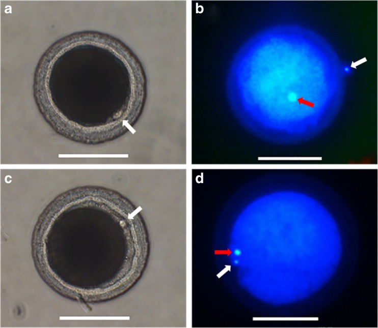
Fig. 4.
First polar body extrusion and chromatin configuration in canine oocytes. a First polar body extrusion in the control group. b Metaphase II (MII) in the control group. c First polar body extrusion in the oviduct cell (OCs) group. d MII in the OCs group. Red arrows MII plate; white arrows first polar body. Scale bar = 100 μm

