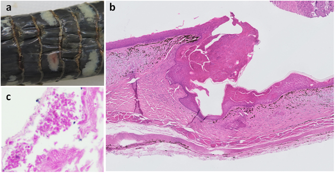
Figure 1.

(a) Macroscopic lesions of snake fungal disease in a grass snake (Natrix natrix) showing thickened, yellow-brown areas mostly at the edges of the ventral scales with irregular margins (case: XT1041-16); (b) Microscopic lesions of snake fungal disease in a grass snake showing epidermal thickening and necrosis and dermatitis (case: X1041-16), HE stain, 50x magnification; (c) Focused area of microscopic lesion showing presence of arthroconidia in grass snake (case: XT804-15), PAS stain, 1000x magnification.
