Figure 1.

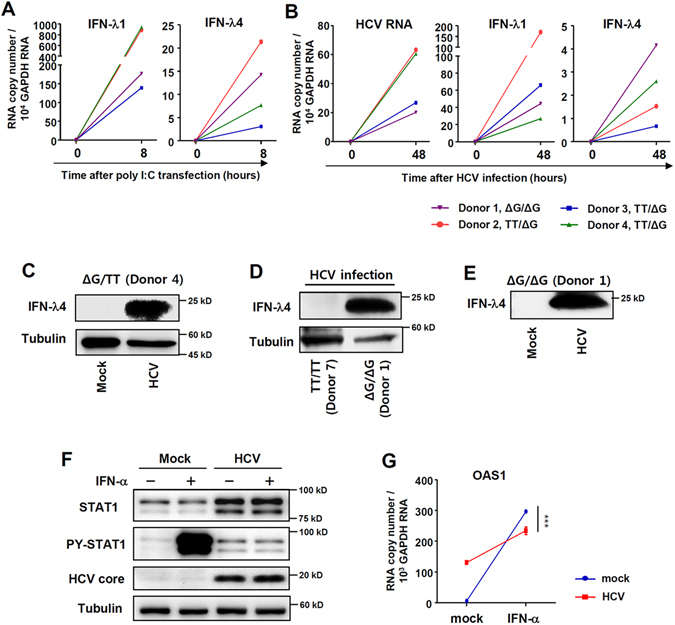
HCV infection results in IFN-λ4 expression and IFN-α unresponsiveness. (A) PHHs from 4 different donors were transfected with poly(I:C) (6 µg/ml). After 8 hours, the cells were harvested, and gene expression was analysed by real-time qPCR. (B) PHHs from 4 different donors were infected with JFH1 HCVcc at 10 MOI. After 48 hours, the cells were harvested, and gene expression was analysed by real-time qPCR. (C) PHHs with IFNL4 ΔG/TT genotype were infected with JFH1 HCVcc at 10 MOI. After 72 hours, the cell lysate was harvested, and protein expression were analysed by immunoblotting. (D) PHHs from two different donors (one with IFNL4 TT/TT genotype and the other with IFNL4 ΔG/ΔG genotype) were infected with JFH1 HCVcc at 10 MOI. After 72 hours, the cell lysates were harvested, and protein expression were analysed by immunoblotting. (E) PHHs with IFNL4 ΔG/TT genotype were infected with JFH1 HCVcc at 10 MOI. After 72 hours, the culture supernatant was harvested, and IFN-λ4 protein expression was analysed by immunoblotting. The data are representative of two independent experiments. (F,G) PHHs with the IFNL4 ΔG/ΔG genotype were infected with JFH1 HCVcc at 5 MOI. After 72 hours, the cells were treated with 100 IU/ml IFN-α for 30 minutes (F) or 6 hours (G). The cells were harvested, and gene and protein expression were analysed by immunoblotting (F) or real-time qPCR (G). Data are presented as means ± S.E.M. ***P ≤ 0.001 (Student’s t-test). All the data are representative of two independent experiments. Full-length blots of PY-STAT1 (F) with multiple exposure times are included in the Supplementary Fig. 9A.
