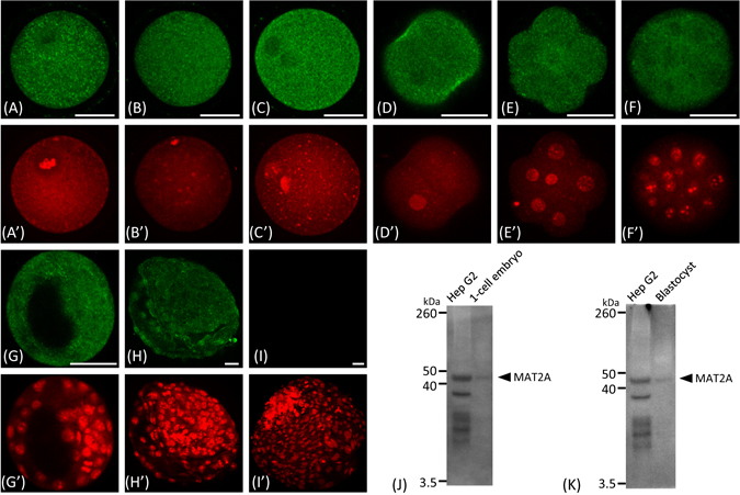
Figure 1.

Immunofluorescence analysis of MAT2A protein in bovine oocytes and preimplantation embryos. Confocal images that transverse at least one nucleus or pronucleus are shown. (A) Immature oocyte, (B) mature oocyte, (C) 1-cell, (D) 2-cell, (E) 8-cell, (F) morula, (G) blastocyst, (H) hatched blastocyst, and (I) negative control in which the primary antibody was omitted from the immunofluorescence of a hatched blastocyst sample. Scale bars represent 50 µm. (A’–I’) Nuclear counterstaining with propidium iodide. (J) and (K) Immunoblotting of 1-cell embryo (n = 120) and blastocyst (n = 120) lysates using the monoclonal MAT2A antibody used in the immunofluorescence and ChIP-seq analyses. Hep G2 Cell Lysate (50 µg protein) was used as a positive control.
