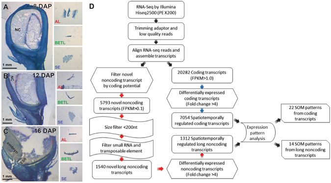
Figure 1.

Schematic of method for transcriptome analysis of developing maize endosperm. (A–C) HistoGene-stained frozen sections from kernels at (A) 8 DAP, (B) 12 DAP, and (C) 16 DAP. Three insets on the right side of each panel show the three tissue types isolated from cryo-sections by free-hand dissection. The tissues surrounded by the red dashed lines and the blue dashed lines correspond to the AL and SE tissue, respectively. The BETL is marked with green dashed lines. NC, nucellus. (D) The bioinformatics pipeline used to identify spatio-temporally regulated coding and long noncoding transcripts at three stages of the maize endosperm development.
