Figure 1.

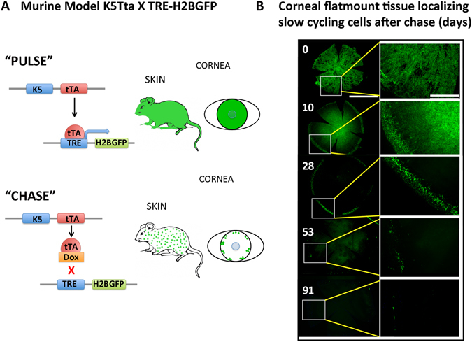
Murine K5Tta × TRE-H2BGFP identifies limbal localized GFP+ cells in corneal wholemount tissue. (A) Tetracycline-inducible (tet-off) double transgenic mouse system drives expression of histone H2B–GFP from the epithelial keratin 5 (K5) promoter (pulse). Dox administration in mouse diet turns off H2B–GFP expression (chase) when proliferating epithelial cells dilute the label between daughter cells by divisions. (B) Corneal flatmount tissue localizing slow cycling cell populations from K5tTA X TRE-H2BGFP mice starting from 0–91 d chase. Low magnification of the entire cornea is displayed on the left panel after 0 (n = 4), 10 (n = 2), 28 (n = 3), 53 (n = 5) and 91 (n = 3) d chase and higher magnifications from the boxed areas on the left panel are shown on the right. GFP+ nuclear staining marks the majority of the corneal epithelium at 0 d chase. At 10 d chase GFP positive nuclei are scattered throughout the cornea, with a distinct lack of GFP+ cells separating the periphery and the central cornea. Low magnification of 28 d chase shows few GFP+ cells in a cluster localized to the periphery. The enlarged image at 28 d shows these GFP+ nuclei at the peripheral base and the limbal region. Low magnification of 53 d chase exhibits fewer cells in the periphery compared to 28 d chase, and the enlargement shows the limbal localization of these cells. At 91 d chase, GFP+ cells become few, but the location of GFP+ cells remain at the limbus as highlighted in the enlarged image on the right panel. Scale bar in B for right panel = 1000 µm and for the left panel images = 400 µm.
