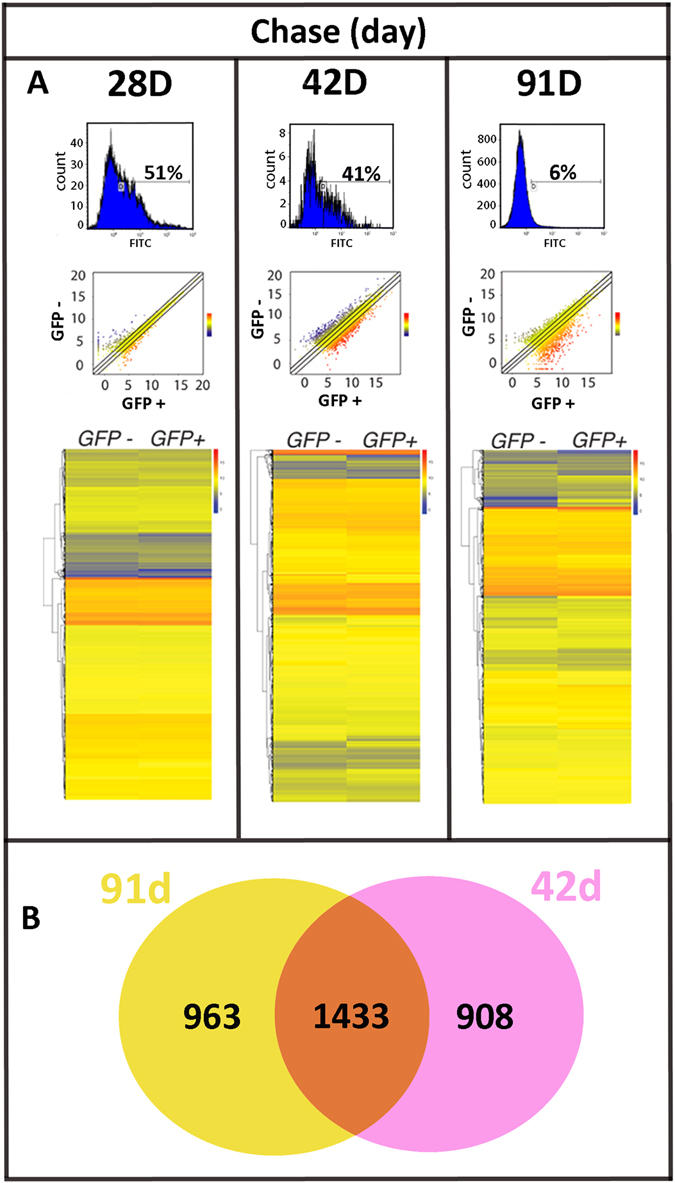
Figure 2.

Differential gene expression between GFP− and GFP+ corneal cells are greater at 42 d post chase and thereafter. (A) FACS analysis of freshly dissociated K5tTA X TRE-H2BGFP corneal epithelial cells demonstrated decreased GFP+ LRC percentages as the chase period was extended from 28 d (51%, n = 5) to 42 d (41%, n = 3) and to 91 d (6%, n = 5). The scatterplot of the RNA-Seq expression data shows the differences in gene expression between GFP+ (x-axis) and GFP− (y-axis) in log2-transformed scale from 28 d, 42 d and 91 d chase. Gene expression change between GFP− and GFP+ is represented as a color (blue, low expression to red, high expression). Differential gene expression increases at 42 d and 91 d chase. The heat map includes 12,597 genes for each chase period. Color differences between GFP− and GFP+ become more apparent at 42 d chase onwards. (B) Venn diagram of significantly differentially expressed genes between GFP− and GFP+ shows there are 1433 genes (orange) shared between 42 d and 91 d chase. There are 908 genes (pink) exclusively differentially expressed at 42 d and 963 genes (yellow) exclusively differentially expressed at 91 d chase.
