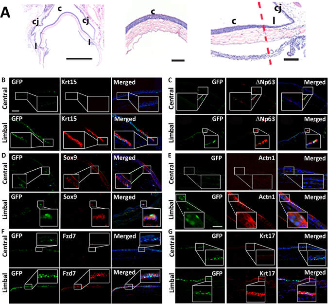
Figure 6.

K5Tta × TRE-H2BGFP+ slow cycling cells co-localize with corneal stem cell markers. (A) Low power haemotoxylin and eosin staining of the adult cornea, limbus and conjunctiva tissue (left), magnified images of the central cornea (middle) and limbal cornea (right). (B–G) Corneal immunofluorescence staining of K5Tta × TRE-H2BGFP sections, co-stained with putative stem cell markers in the central (upper panels) and limbal cornea (lower panels). (B) At 35 day chase (n = 3) few lowly expressed GFP+ cells were found in the central cornea while Krt15 (red) antibody staining is not expressed. At the limbus, there is high GFP+ LRC staining at the limbal and periphery basal layer, including to one cell layer above in some areas with weaker expression. Krt15 and few GFP+ cells co-localize in limbal cells as shown in the merged panel. (C) In the central cornea, GFP+ LRCs at 53 d chase (n = 3) and ΔNp63 (red) labeled staining was absent. At the limbus, GFP+ LRCs were localized to 2–3 cells at the limbus and ΔNp63 expression was limbal and basal specific (nuclear expression). The merged image shows the co-localization of GFP and ΔNp63. (D) GFP+ LRCs at 53 d chase localized to a few cells in the central cornea (boxed) while Sox9 (red) marked only basal epithelial cells (n = 6). The merged image co-localizes them together. At the limbus, GFP+ LRCs co-localized with Sox9. (E) GFP+ LRCs from 53 d chase were not detected and Actn1 was devoid of staining in the central cornea. At the limbus, few GFP+ cells were visible and co-localized to cytoplasmic Actn1 staining (n = 4). (F) GFP+ LRCs cells at 35 d chase are strongly expressed in the basal and some suprabasal layers of the central cornea, while some weak staining was also observed in cells of the suprabasal layer. Fzd7 staining in the central cornea is devoid of any expression, while at the limbus, the expression is basal and co-localized to GFP+ LRCs (n = 2) (G) GFP+ LRCs are strongly expressed in the basal and some suprabasal layers of the central cornea, while some weak staining was also observed in cells of the suprabasal layer. Krt17 staining was absent in the central cornea, but co-localized with GFP+ LRCs at the limbus (n = 3). Scale bar in A = 200 µm for (A–C,E,F); in D scale bar = 50 µm.
