
Figure 4.
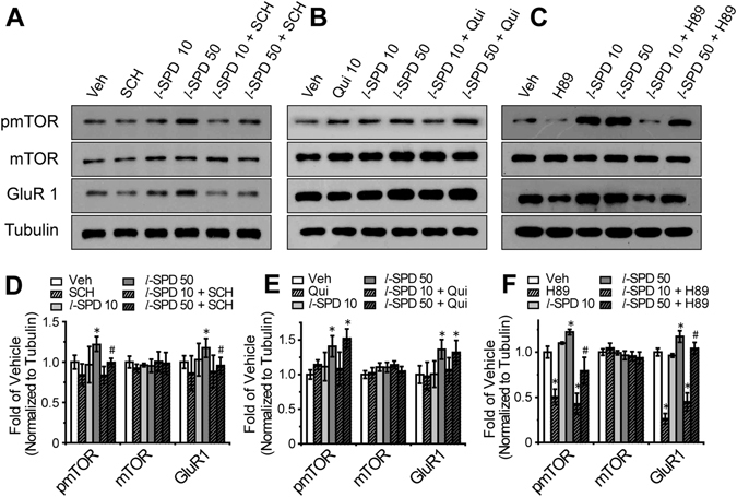
l-SPD selectively activates mTOR signaling by D1R agonism rather than D2R antagonism. One week cultured primary cortical neurons were incubated with drugs for 4 hours. (A) Western blot images and (D) quantification analysis of pmTOR (Ser 2448), mTOR, GluR1 in the treatment of vehicle, l-SPD (10 µM or 50 µM), SCH23390 (10 µM) and SCH23390 (10 µM) with l-SPD (10 µM or 50 µM). (B) Western blot images and (E) quantification analysis of pmTOR (Ser 2448), mTOR, GluR1 in the treatment of vehicle, l-SPD (10 µM or 50 µM), Quinpirole (10 µM) and Quinpirole (10 µM) with l-SPD (10 µM or 50 µM). (C) Western blot images and (F) quantification analysis of pmTOR (Ser 2448), mTOR, GluR1 in the treatment of vehicle, l-SPD (10 µM or 50 µM), H89 (10 µM) and H89 (10 µM) with l-SPD (10 µM or 50 µM). The β-tubulin were determined as reference protein. *p < 0.05, compared to the vehicle treatment group. # p < 0.05, compared to the 50 µM l-SPD treatment group.