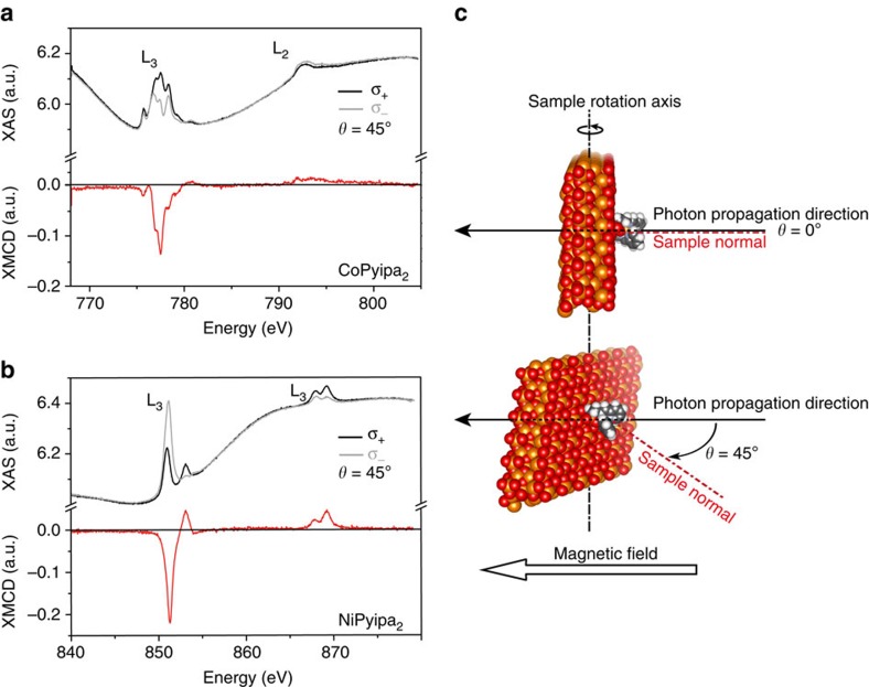
Figure 4. XAS/XMCD spectra of a monolayer of Co- and Ni(Pyipa)2.
(a) Cobalt, and (b) Nickel L2,3 edges XAS (black and grey line) and XMCD (red lines) spectra recorded at T=2 K, and θ=45° using left (σ+) and right hand (σ−) circularly polarized light in 6.5 T field; (c) schematic representation of the measurement geometry.

