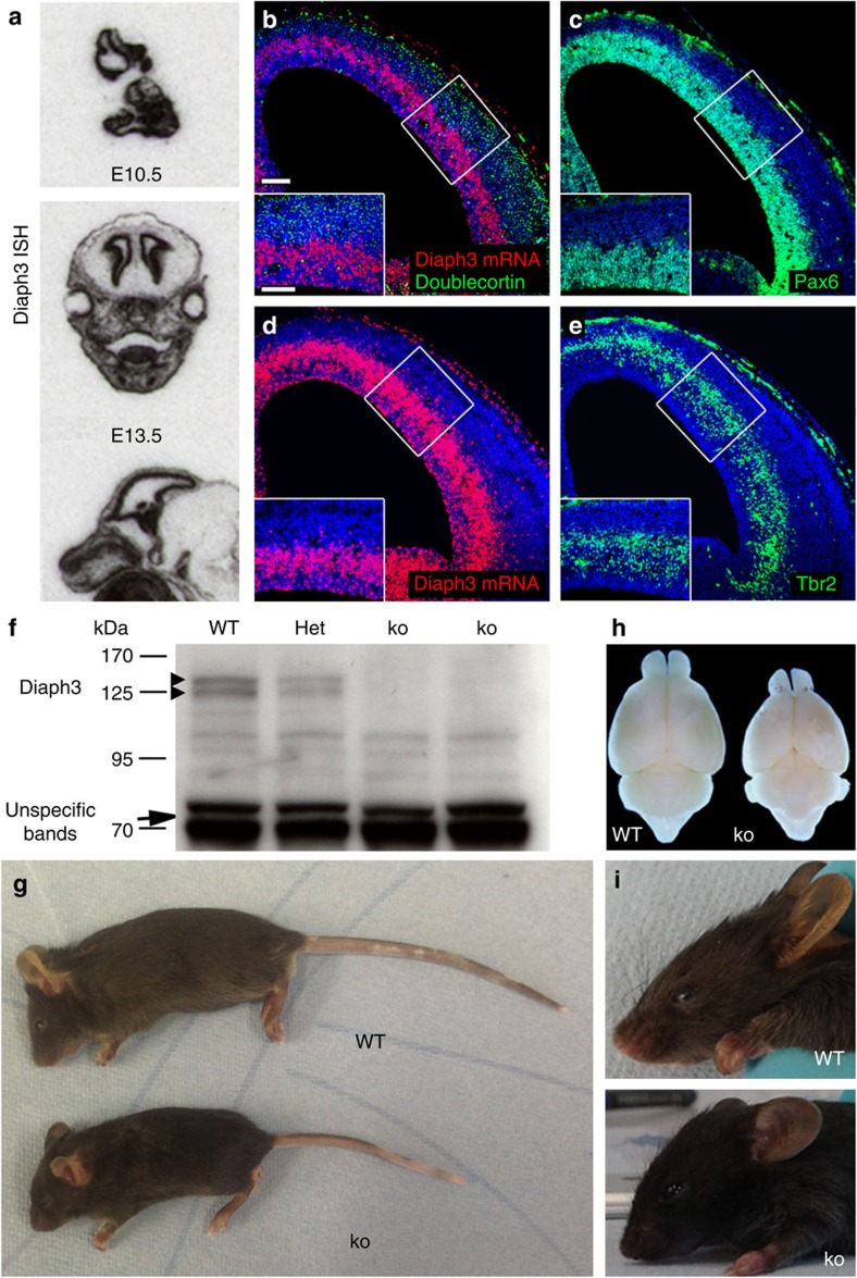
Figure 1. Generation of Diaph3 mutant mice.
(a) Sections of E10.5 embryos and E13.5 heads, hybridized with a Diaph3 radiolabelled probe. The mRNA signal is associated with the telencephalic VZ where the progenitor cells reside. (b–e) Coronal sections of E13.5 brain hemispheres hybridized with a fast red-labelled RNAscope probe. The signal localized mostly in the outer part of the VZ (b,d). No signal was found in committed cortical neurons stained for Doublecortin (green, b). (c,e) Sections adjacent to those illustrated in b,d, stained for Pax6 (c) and Tbr2 (e). Scale bars, 100 μm and 50 μm in the insets. (f) Western blotting of E13.5 embryo lysates from WT, heterozygotes (Het) and mutant (ko) littermates. The absence of Diaph3 protein doublet in Diaph3 ko mice (arrowheads) is noteworthy. Nonspecific bands at 70 kDa provide a loading control (arrow). (g–i) Comparison between Diaph3 WT and ko littermates at P25. The small body size (g), small brains (h) and facial deformities (i) in Diaph3 ko mice are noteworthy. ISH, in situ hybridization.

