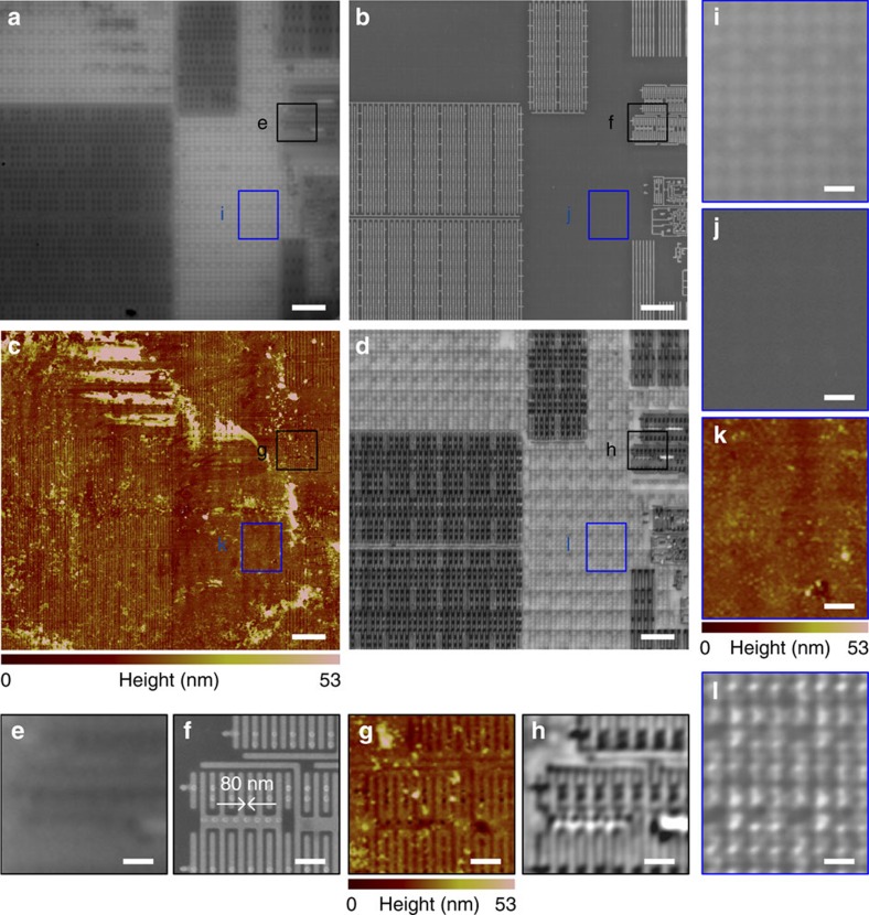
Figure 3. CPU sub-surface structure imaging in constant-height scanning mode.
Comparison of (a) a conventional microscope mounted with a × 100 (numerical aperture=0.8) objective, (b) SEM, (c) AFM and (d) the SSUM for the observation of CPU sub-surface structures beneath a 10±2 nm (measured by AFM)-thick optically transparent film preventing AFM and SEM detection, which are denoted by i–l. (e–l) Local zoomed areas that correspond to the marked areas in a–d. Scale bars, 5 μm (a–d); 1 μm (e–l).

