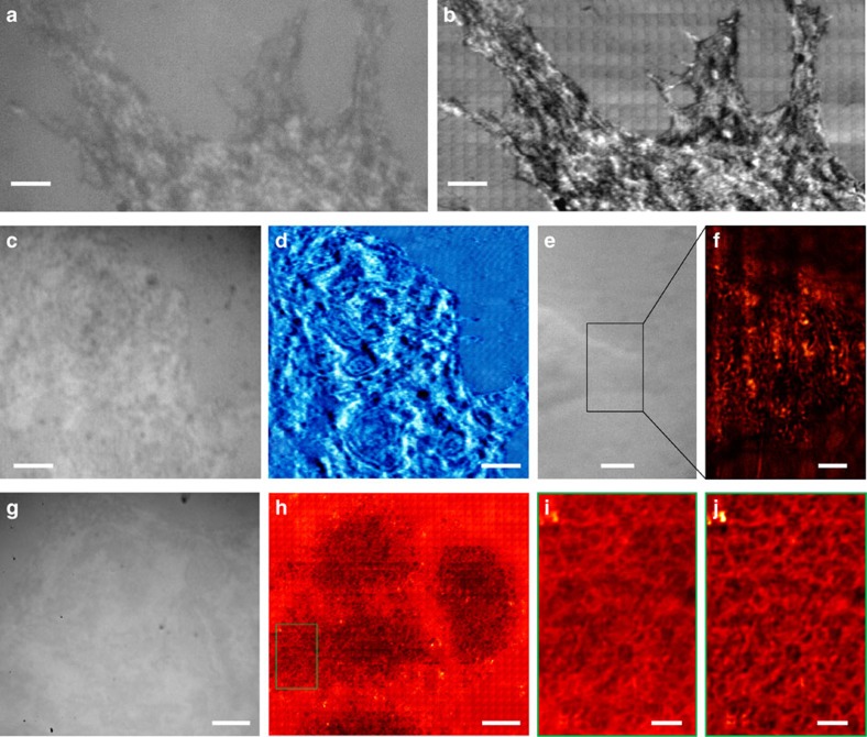
Figure 5. Non-invasive observation of cells in white-light mode.
A C2C12 cell was imaged using (a) a traditional optical microscope or (b) SSUM. A video recorded while scanning a C2C12 cell is provided as Supplementary Movie 2. MCF-7 cells were observed (c,e,g) without and (d,f,h) with the aid of the microsphere superlens. A × 100 (numerical aperture (NA)=0.8) objective was used in a,b,g and h, and a × 50 (NA=0.6) objective was used in c–f. (i) Local zoomed area of the marked area shown in h. (j) After using a band-pass filter algorithm of i. Scale bars, 6 μm (a,b); 10 μm (c–e,g,h); 3 μm (f); 2 μm (i,j).

