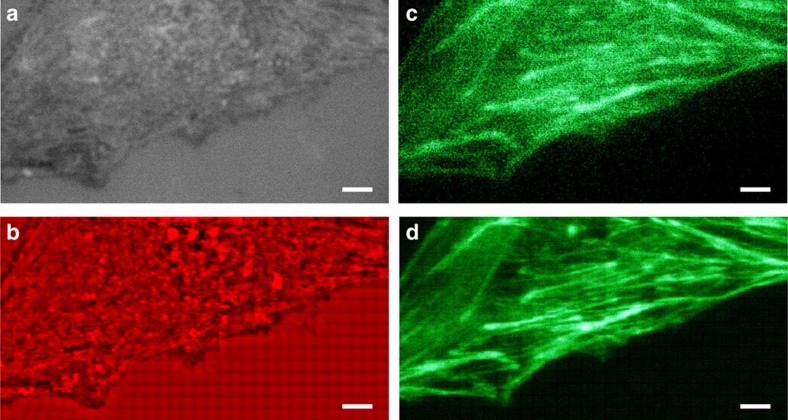
Figure 6. Non-invasive white-light and fluorescence microscopy of a C2C12 cell.
(a,b) White-light and (c,d) fluorescent imaging of a C2C12 cell (a,c) without and (b,d) with the enhancement of a 56 μm-diameter microsphere superlens. A × 100 (numerical aperture=0.8) objective was used in these experiments. For fluorescent imaging, the sample was labelled by Alexa Fluor 488-phalloidin to observe actin filaments. Scale bars, 5 μm.

