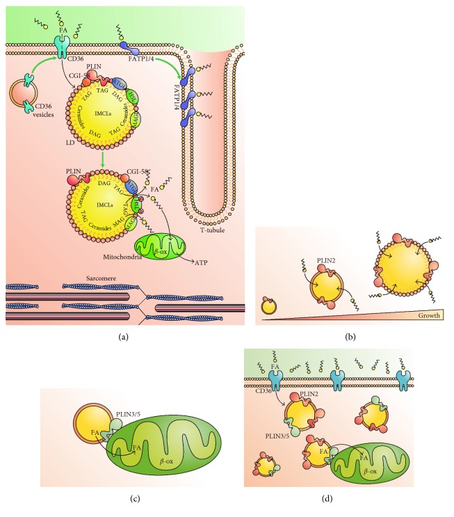
Figure 1.
FA flux in skeletal muscle and role of perilipins. (a) Schematic representation of FA uptake and deposition in lipid droplets (LD). FA uptake is mediated by FAT/CD36, located in the plasmalemma. FATP1/4 are located in the plasmatic membrane and cooperate with FA intake and metabolism. Movement of CD36 to the plasmatic membrane and an increment in FATP1/4 can be triggered by insulin or contraction (green arrow, see text for details). Once inside the cell, FAs can be accumulated in LDs as acyl-glycerides (TAG, DAG). Perilipins (PLINs) coat the LD membrane, along with the lipases ATGL and HSL and the coactivator CGI-48. In response to contraction (green arrow), ATGL activity increases, as a result of dissociation from PLINs, while HSL is activated by PKA-dependent phosphorylation. This leads to increased FA flux to cytosol and mitochondria, undergoing further β-oxidation (β-ox) and ATP synthesis. (b) Main function of PLIN2 in skeletal muscle. PLIN2 coats LD and promotes FA intake, leading to increased size of LD. (c) PLIN3 is part of LD mitochondria contact sites, promoting efficient transfer of FA from LD to mitochondria for oxidation. (d) In cases of excess plasmatic FAs, such as obesity and T2DM, FAs uptake into skeletal muscle is increased. Higher levels of PLIN2 promote an increase in both size and number of LDs. Furthermore, increased levels of PLIN5 foster FA metabolization inside mitochondria.

