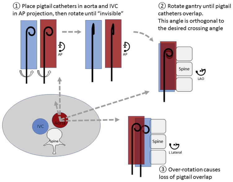
FIGURE 7. Finding the Optional Transcaval Projection Angle Without Using Computed Tomography.
The lower left figure depicts an abdominal cross-section at the level of the intended transcaval crossing, with a typical relationship of inferior vena cava (IVC), aorta, and spine. Pigtail catheters are placed in the IVC and the aorta and rotated in the anteroposterior projection until the pigtail curve is no longer visible. Then rotate the fluoroscopy gantry left-right until the 2 pigtail catheters overlap. A small-volume contrast injection can confirm appropriate positioning. Over-rotation also is depicted, at which point pigtail catheters no longer overlap. AP = anteroposterior projection angle; LAO = left anterior oblique.

