Figure 3.
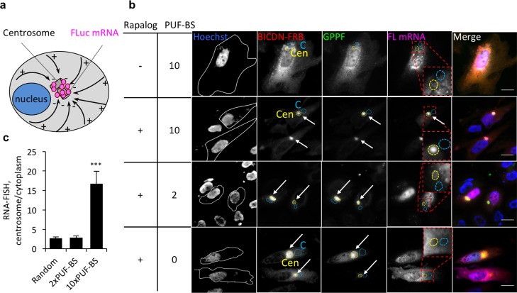
Transport of reporter mRNA to the proximal ends of microtubules in HeLa cells. (a) Schematic overview of BICDN-mediated mRNA transport. Microtubules are represented as black solid arrows pointing from their (−) to (+) ends. (b) Transport of FLuc mRNA to the perinuclear region by BICDN-FRB and the PUF construct GPPF. Hoechst, nuclear stain. White lines, cell outlines. Quantified areas: C, cytoplasm (light blue dashed lines); Cen, centrosome (yellow dashed lines). PBS, PUF-binding sites. Enriched spots indicated with arrows. Scale bar: 20 μm. (c) Quantitation of mRNA transport to the perinuclear region. RNA-FISH intensity at the region coinciding with the brightest fluorescent region of BICDN immunofluorescence was normalized to an adjacent distal region of the same area. n = 20 cells in 3 biological replicates. ***P < 0.001. Mean ± SEM.

