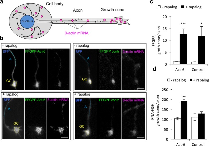
Figure 4.
Transport of endogenous β-actin mRNA to axonal growth cones in primary neurons. (a) Schematic of endogenous mRNA transport in hippocampal neurons. Microtubules are represented as black solid arrows pointing from their (−) to (+) ends. (b) Effect of FFGPP-Act-6 or FFGPP-control on abundance of β-actin mRNA in axonal growth cones. FFGPP-act-6 PUF domains recognize β-actin mRNA and FFGPP-control PUF domains recognize unrelated 5′UUGAnAUA3′. BFP was transfected to track the neuron outline. Quantified regions: GC, growth cone (yellow dashed lines); A, axon (light blue dashed lines). Scale bar: 5 μm. (c) Quantitation of FFGPP redistribution. FFGPP fluorescence in a growth cone was normalized to fluorescence measured along a 10–20 μM line following the axon approximately 10–20 μM away from the growth cone. (d) Quantitation of β-actin mRNA in axonal growth cones in the presence of FFGPP-act or FFGPP-control. n = 20 cells in 3 biological replicates. Mean ± SEM. ***P < 0.001. **P < 0.01. *P < 0.05.

